
Biomechanical Imaging with PDE-Constrained Optimization
At SCOREC, I implemented adjoint-based PDE-constrained optimization algorithms for large-scale simulation with FEniCS in Python. I also developed Split Bregman methods for L1-regularized nonlinear PDE-constrained optimization and applied them to recover material parameters in biological tissues from measured displacement fields.
Applications included OCT and ultrasound-based elasticity imaging, adaptive meshes, spatial domain decomposition, and continuation strategies for linear and nonlinear tissue models.
 Mesh adaptivity of a stiff inclusion in a homogeneous tissue-mimicking phantom.
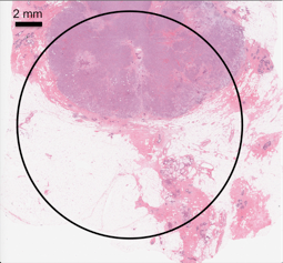 H&E histology of a malignant breast tumor tissue. The black circle is the region of interest.
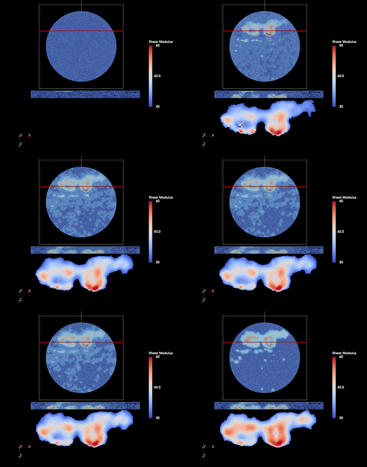 Mesh adaptivity and shear modulus distribution of malignant breast tumor tissue ex vivo. Both elliptical tumor tissues have soft necrosis centers.
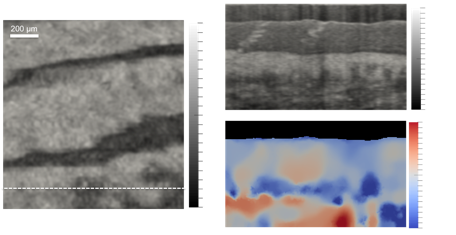 OCT en-face image of fingertip tissue. The B-scan and recovered shear modulus distribution show the triple-layer tissue structure and sweat glands.
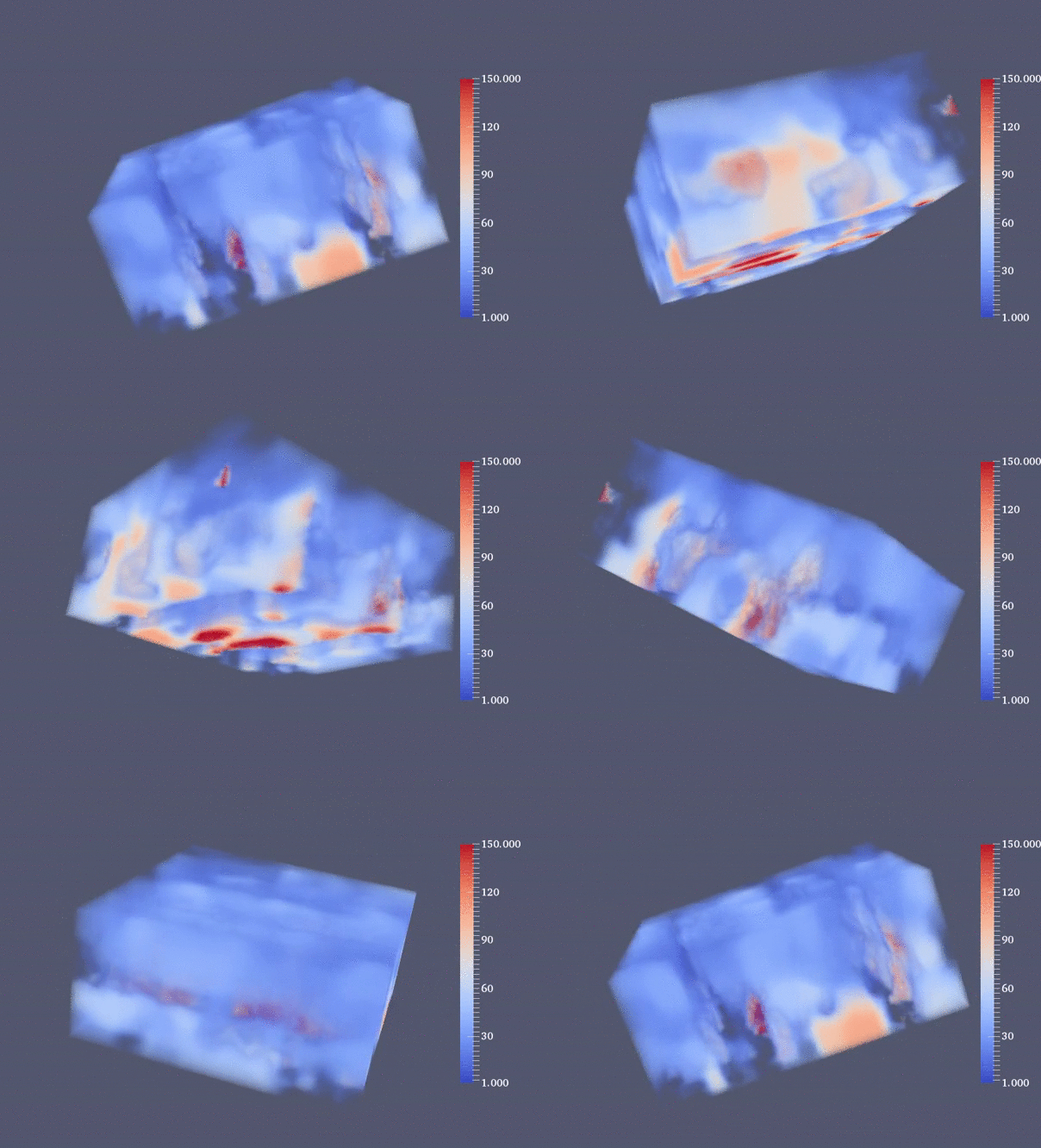 3D shear modulus distribution of fingertip tissue in vivo. The three stiff ridges are more-compressed tissue under bulged fingerprints.
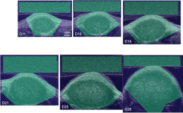 Tumor evolution on days 11, 15, 18, 21, 25, and 28. The ultrasound B-mode regions of interest are shaded in green.
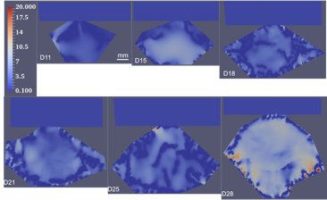 Recovered shear modulus distributions for the same tumor-evolution sequence.
Related publications:
- Dong, L., Wijesinghe, P., Dantuono, J. T., Sampson, D. D., Munro, P. R. T., Kennedy, B. F., & Oberai, A. A. (2016). Quantitative compression optical coherence elastography as an inverse elasticity problem. IEEE Journal of Selected Topics in Quantum Electronics, 22(3), 277-287.
- Dong, L., Wijesinghe, P., Sampson, D. D., Kennedy, B. F., Munro, P. R. T., & Oberai, A. A. (2019). Volumetric quantitative optical coherence elastography with an iterative inversion method. Biomedical Optics Express, 10(2), 384-398.